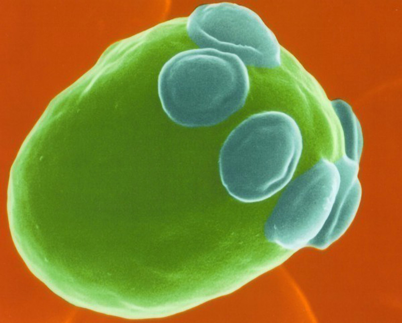
A colorized scanning electron microscope image of a single cell of Sacchromyces shows the budding scars in blue where the cell has reproduced asexually.

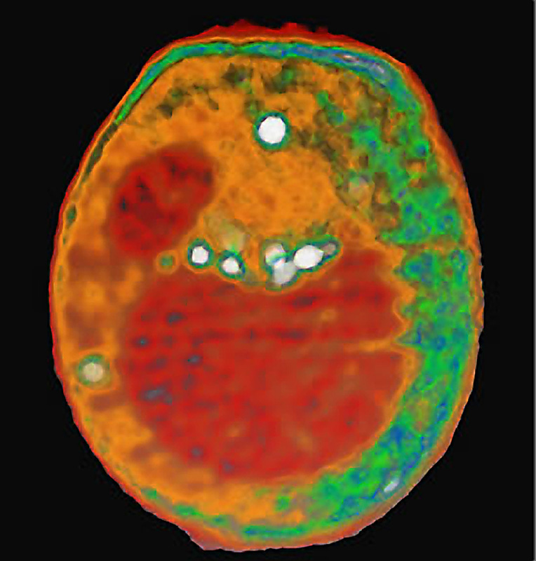
This x-ray image of a yeast cell was taken at the Advanced Light Source at Lawrence Berkeley National Laboatory. Its Internal organelles are color-coded according to x-ray absorption. The nucleus is the smaller red sphere and the large red sphere is a vacuole. Lipid droplets are shown in white and cytoplasmic structures are shown in either orange or green.

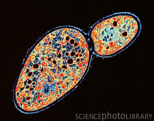
Budding yeast cell (Saccharomyces Cerevisiae)


